Making Innovative X-Ray Imaging Accessible Everywhere - Interview with KA Imaging
2022/02/14
The invention of X-ray imaging revolutionized medicine by providing a means for noninvasive disease diagnosis and monitoring. We interviewed the co-founders of KA Imaging, a startup that is re-revolutionizing the field with portable dual-energy X-ray technology.
Interviewer: Suzu Kitamura
Company Profile: KA Imaging
KA Imaging is a spin-off startup from the University of Waterloo, specializing in the development of novel X-ray imaging technologies and systems. Based in Canada, their vision is to make innovative X-ray imaging accessible everywhere.
Amol Karnick
President and Chief Executive Officer
Amol Karnick holds a Bachelor's degree in Engineering from the University of Waterloo and a Masters in Electrical Engineering from McGill University. Since entering the medical imaging industry, Amol has accumulated 20 years of experience at numerous startups and large medical corporations.
Dr. Karim S. Karim
Chief Technology Officer
Dr. Karim S. Karim is a professor at the University of Waterloo and has been developing innovative X-ray imaging technologies for over 20 years. He is the co-author of over 250 publications and 80 patents. Dr. Karim is a serial entrepreneur with experience supporting two startups and founding two companies.
-- Can you tell us a bit about yourselves and about your company?
- Amol
Karim and I met back in 2011 when I was working as an executive-in-residence at Communitech. Karim joined through his University transfer program. We met and had a conversation, but then I left and he went away on a sabbatical. Upon his return he said “I’ve got this great technology in my lab, would you like to work with me?” I looked at the technology and agreed. At first, we kept the technology incubated at the University of Waterloo until 2015, when we raised our first round of capital and created KA Imaging.
- Karim
KA Imaging has a very close connection to the University of Waterloo. It’s the largest engineering university in Canada, and it’s also the home of the Blackberry cellphone. It has very strong technical capabilities. Actually, the University of Waterloo has 20 to 30 years of experience building X-ray sensors. A lot of our research employees have come from the university, so we have a very strong technical staff.
-- Please tell us more about the technology you are working on at KA Imaging.
- Amol
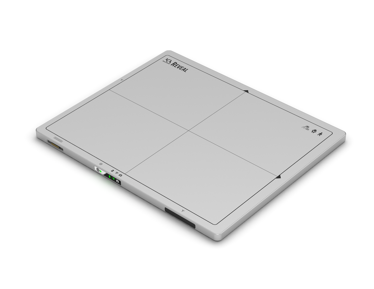
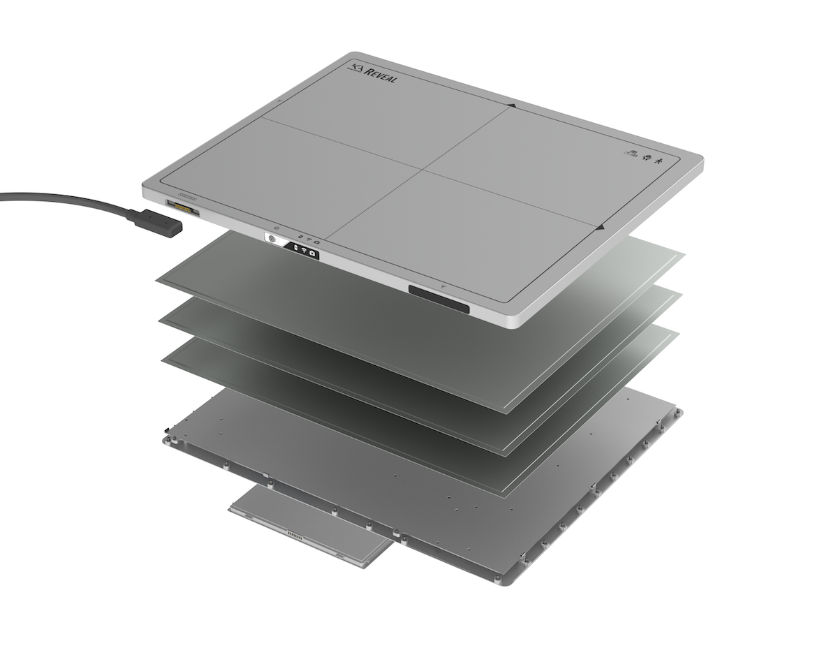
Our goal is to make better diagnostics accessible globally. We have two X-ray detector technologies in house. One is the Reveal technology, which is a multilayered detector. The other detector is called BrillianSE. It’s a very high resolution CMOS* selenium detector that we use in our microCT called Insight.
*CMOS is a type of metal-oxide-semiconductor field-effect transistor (MOSFET) fabrication process which consumes less energy while allowing high speed transmission.
- Karim
We are leveraging the most cutting edge, advanced technology in X-ray, including spectral X-ray, phase contrast X-ray and dual energy X-ray. The portability of our product is key – we are the first and only detector in the world that can do portable dual energy imaging. The technology we have developed is protected by 80 global patents, 22 of which are unique. It is very rare in the detector space to have patented technology. Aside from our hardware, we also have a number of algorithms to leverage our X-ray technology. These algorithms generate the software platform, which together with our hardware platform, gives the best image quality possible.
-- What differentiates your technology from that of competitors?
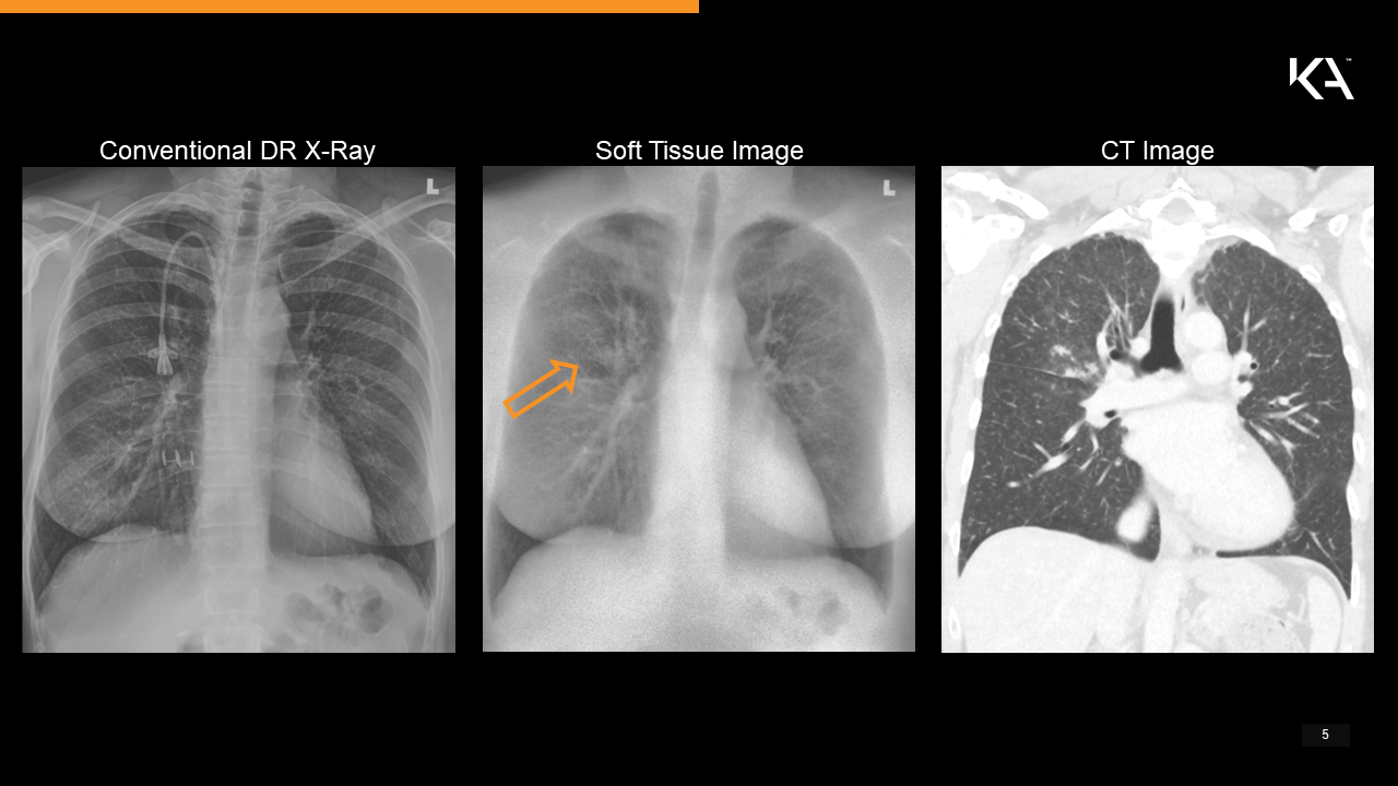
- Karim
The main difference is our dual energy technology. Dual energy X-ray has been around in the market for 30 years, but it always requires changes to be made to the existing conventional X-ray system in place. It usually uses more radiation, requires different imaging techniques, is not portable, or cannot take lateral images. There is always work required to integrate the dual energy technology. We are the only supplier of dual-energy X-ray that doesn’t change anything. We use the same radiation, the same clinical technique, and our technology can be used in a portable form factor, and we can take lateral and oblique images. We can do everything that regular X-ray can do, and, in addition, for every single X-ray exposure, our customer gets three images – regular, soft tissue, and bone. Our advantage is that we have taken dual energy from being a niche to being mainstream. Now, everyone can get dual energy with every scan they do, whether it be a scan for the chest, head, abdomen, extremities, or even for a pediatric patient.
Our biggest secret is that while everybody else is using two exposures – a low energy exposure and a high energy exposure – we use a three-layer sensor. With the two-layer model, one always has a tradeoff between image quality and dose efficiency. The advantage of our design is that the third layer allows us to get 100 % dose efficiency, the best dose compared to any existing digital X-ray detector. Also, our X-ray detector is portable because all of the dual energy spectral separation happens inside of the detector. The source of the X-rays can be any source, no additional filter is necessary, nor any special changes.
-- Where is your technology being used? How does it get installed?
- Karim
We’ve been cleared in Canada and the United States by the FDA as a general radiography X-ray detector. We are also approved and have evaluation units in Australia, Philippines, Malaysia, Indonesia and Pakistan. People are using our technology in regular clinical practice.
- Amol
We had a clinical trial at the University Health Network in Toronto for pneumonia and COVID-19, and are having a trial at Grand River Hospital for lung cancer. Our X-ray detector can upgrade any existing system without changing anything, and it takes us less than half a day. We can just visit a site and change the detector to ours,the process itself takes less than an hour. The rest of the time is spent training the personnel on how to acquire images, and the acquiring process is very similar to the conventional process. By contrast, our competitors would have to do a full room upgrade, which can take up to a week to finish.
-- What other applications do you foresee for KA Imaging’s X-ray technology?
- Karim
The other applications for this technology other than medical include security, industry, veterinary, and electronics. For security, you can use it as a portable scanner for baggage in airports or train stations. This kind of technology can give you dual energy images, which can help better identify metal and plastic. It can also be applied in non-destructive tests for aeroplane wing inspections. Aeroplane wings are often made out of composite materials including titanium which requires careful inspection at times. This technology would allow you to separate titanium from composites. Another application is in veterinary medicine; for example, looking at the legs of race horses for bone bruises, or finding foreign objects in cats and dogs. The last potential application is electronics. Dual energy is very useful for separating metals and plastics, for example, on a dual circuit board. So you could use it for inspection of televisions, tablets, or computers.
-- What do you think will be your main challenges going into the future?
- Karim
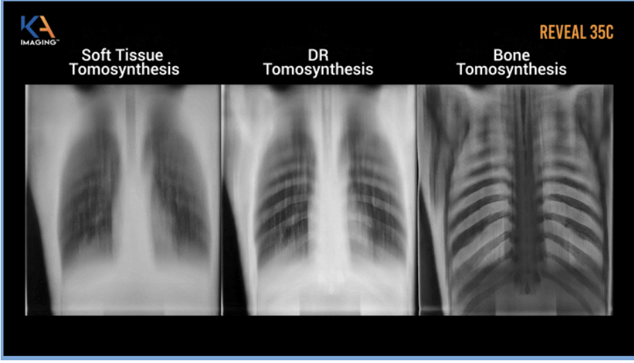
The technical challenges we are continuing to solve include building a flexible (more rugged) version of our detector. Also, we’ve released the world’s first images for single exposure tomosynthesis using dual energy, and we’re looking to create dual energy that’s portable with tomosynthesis*. There are quite a few interesting challenges and opportunities that exist for our three-layer design. We’re looking at using it for uses other than chest X-ray, such as mammography, fluoroscopy, and of course pediatrics.
*Tomosynthesis is a novel type of digital X-ray mammogram that creates 2D and 3D images of breast tissue, improving the ability of mammography to identify early breast cancer.
- Amol
We’re actively seeking investment for our Series B round. We’re starting the round now and we’re looking to raise US$ 25 million.
-- What are you looking for in potential partners? What unique opportunities do you see in the Japanese market?
- Amol
We are looking for partners that are system integrators and people who are selling tomosynthesis systems on the market. We would like for them to work with us so we can integrate our detector and improve tomosynthesis, so that patients can have the best outcome.
Furthermore, what we would like is the opportunity to be connected to hospital systems in Japan. What we need even before distributors and integrators is the opportunity to talk to hospitals and install our technology, so we can use it locally and collect data.
Regarding unique opportunities in Japan, to my understanding, almost every adult in Japan has an annual chest X-ray. We think this is a great opportunity for us because if you look at the healthcare side of it, our technology improves chest X-ray imaging. With our technology, you can spot potential lung cancer, pneumonia, or cardiac disease earlier – in fact, identifying calcified coronary arteries is something that current X-ray cannot even do. Because of the high per capita usage of X-ray in Japan and the annual screening of a large number of adults, we think Japan is a fantastic market. In the long run, our technology will improve the capability to find disease early.
We would like to express our sincerest thanks to Amol and Karim for taking part in this interview.
KA Imaging is participating in the Plug and Play Japan Health accelerator program for its fifth batch. Learn more about the company and their technology from the link below.